2022年3月20日星期日

Wiseye PLX119C Mobile C-arm System with Upgraded FPD

 
Large view
Small footprint
TECH
CONTRIBUTES

HEALTH

Upgraded FPD
Revealing more anatomical details

Equipped with 30cm*30cm large size dynamic FPD, imaging area has been increased by 100%compared with the conventional FPD.

4MP resolution image combined with low signal-noise ratio reveals tiniest anatomicaldetails.

Double the image size
Decrease exposure times

5 lumbar vertebrae can be photographed within one image by a 30cm x 30cm FPD, largely decreasing exposure times caused by insufficient image FOV, making it easier for surgeon to locate lesions and to make surgical plan in the procedure like pedicle screw fixation or procedure with strict imaging demand.

lmage taken by 30cm x 30cm FPD can basicly cover the entire pelvic floor. In the surgery of bilateral-pevicfractures or pelvic ring internal fixation, all the fracture site can be revealed by a single exposure, which significantly improves overall efficiency.

Clinical image

Clinical application

lmage with high resolution can clearly reveal articular structure, helping surgeons to better locate the target

Flexible mechanical movement realize easy positioning for different type of trauma surgery, providing comfortable handling experience.

upgraded FPD present more details of vertebrae, helping surgeons to better making surgical plans.

Multiple radiation protection decrease overall exposure times during minimally invasive procedure.

Low dose management
Multiple protection

Leakage radiation in the loading state of X-ray tube is far more lower than CFDA and FDA standard, providing better protection from the X-ray source.

X-ray generated in high frequency pulse wave can decrease 50% of radiation dose compared with a continous fluoroscopy. This particular technology has a positive effect on weaking the motion artifact.

Real time DAP meter displayed on image.Easy for monitoring radiation level.

Optimized and adjustable exposure parameter created by intelligent hardware and algorithm provide a friendly examination scenario.

Allowing radiologist to preview and to adjust exposure area before exposure, reducing unnecessary radiation.

Decreasing radiation level by absorbing soft X-ray.Ensuring high quality image for precise diagnosis and treatment.


Medical Diagnostic X-Ray Equipment; Sharp Image Support Your Accurate Diagnose

 The PLD6500 high frequency digital x-ray equipment can be applied to satisfy the radiographic needs of different body parts: head, chest, abdomen, waist,thorax, pelvis, and limbs.The PLD6500 is also capable of serving different positions: lay decubitus,normotopia, side position,etc.It is designed as a fixed digital x-ray machine, though it can also be compacted into limited spaces, like buses and trucks.


Sharp Image Support Your Accurate Diagnose

63KW generator makes it different,the generator assures the best image in every operating condition along with reduced patient close and extend x-ray tube lifetime.

PLD6500-2

● The PLD6500 high frequency digital x-ray equipment can be applied to satisfy the radiographic needs of different body parts: head, chest, abdomen, waist, thorax,pelvis, and limbs.
● The PLD6500 is also capable of serving different positions: lay decubitus, normotopia, side position, etc.
● It is designed as a fixed digital x-ray machine, though it can also be compacted into limited spaces, like buses and trucks.

DR DETECTOR

Flat panel detector improvse your workflow,exam speed and comfort with efficiency
A-si detector with CSI screen,excellent spatial resolution
Active area:17” x17″

X-RAY SOURCE ASSEMBLY

our generator provide the right amount of penetration, reducing exposure times and retakes.
Automatic exposure control(AEC) enables auto selection of radiographic factors, saves time, eliminates retakes, increases diagnostic capability and lowers the radiation dose.(Optional)


Image acquisition workstation DRA20l

Computer system
Processor: Intel Core Quad Core Processor i5-6500 (3.20 GHz)
Memory: 4 GB DDR3L
Hard disk: 1T SATA(7200rpm)
ptical drive:DVD-RW disc burner


2022年3月9日星期三

What are the advantages of the FPD C-arm System

 The FPD C-arm System acts as the surgeon’s eye and is the device used for imaging during surgery. They are widely used in interventional radiology for minimally invasive surgery to guide doctors in accurate positioning during surgery. Due to its low dose and small footprint, it can be flexibly used in orthopedics, surgery, gynecology and other departments in hospitals.

The mobile C-arm can be divided into a flat C-arm and FPD C-arm System according to different imaging systems. So, what is the difference between the two?


In principle, the FPD C-arm System is composed of a reinforcing tube, a tube container, a power supply, an optical system and a bracket. X-rays pass through the human body, form an X-ray image on the input screen of the intensifier tube, and become visible light on the input screen. This visible light passes through the photocathode connected to the input screen, and photoelectrons are emitted from the photocathode. Under the action of the electronic lens formed by the anode, a reduced electronic image is formed on the output fluorescent screen after focusing acceleration. The electronic image is converted into a visible light image through the output screen, and the light image on the output screen is sent to the TV CCD by the optical system, and the image can be displayed on the monitor. .

The flat C-arm is composed of an amorphous silicon layer with a photodiode function as a scintillation layer, and a thin film transistor TFT arranged in an array element manner. X-rays pass through the human body, and the scintillator layer converts the X-ray photons into visible light, and then the amorphous silicon photoelectric layer converts them into electrical signals. Then the scanning controller reads the electrical signals and converts them into digital signals. The digital signal information is displayed on the monitor. show.

Compared with the FPD C-arm System, the flat C-arm has obvious advantages: the image is more stable and clear, the field of view is better, and the software algorithm is more advanced. Compared with the shadow-increasing C-arm, the imaging principle of the flat C-arm avoids the loss in signal conversion, improves the utilization rate of X-rays, greatly improves the imaging speed, and takes less time to obtain the required image information. The dose and radiation time are also reduced.

With the further development of related technologies of flat-panel detectors, the current mobile C-arm is gradually being upgraded from an image intensifier to a digital X-ray flat-panel detector, breaking the limitations of traditional image intensification imaging distortion, signal loss and service life. Lower dose, larger effective imaging area, and higher imaging quality. The dynamic flat C-arm will become the standard equipment for clinical application of minimally invasive interventional fluoroscopic imaging in orthopaedics and pain, making medical diagnosis more accurate

Advanced digital imaging technology——PLX7100A HF Mobile Digital C-arm System Flat Panel Detector

 PLX7100A HF Mobile Digital C-arm System Flat Panel Detector

Advanced digital imaging technology. Sharp image

  • Full view FPD, outstanding image quality and large field of view coverage evenduring image rotation.
  • High dynamic field of view, achieve the optimal image of human body’s soft tissueand skeletal tissue.
  • A-si detector with CSi screen, excellent spatial resolution.


  • Perfect digital imaging chain makes the routine operation easily as follow:

    • Orthopedics department
    • General surgery
    • Orthopedic surgery
    • Traumatology department
    • Urinary surgery
    • Spine surgery
    • Pain surgery
    • Gastroenterology department
    • Gynecology


    • A high performance imaging workstation and image processing system serves you a convenient interface for integration with network.
    • Work-list registration for seamless integration in your hospital environment.
    • Dicom 3.0 network interface for integration with PACS or RIS.
    • CD burning for easy image transfer.
    • Multi-storage methods for easy image transfer.
    • C-arm widely vertical movement range meets various height demand of operation table.
    • Longer orbit sliding range brings you more visible area.
    • C-arm back and forward movement can make non-central parts be covered easier.
    • SID can be adjusted by detector movement to expand exposure area, bring you outstanding image and more operation space.
    • Simultaneous movement in 5 axes.
    • Rapid motorized control, meet accurate position demands.

What are the clinical application effects of mobile C-arm?

 According to the results of the seventh national census, my country's population aged 60 and above has reached 264 million. It is expected that this number will exceed 300 million during the "14th Five-Year Plan" period, and my country will enter the stage of moderate aging from mild aging. In order to protect the basic life of the elderly, many policies and measures have been introduced. Among them, the focus is on the aspect of geriatric medical care, and by increasing the medical level and facilities of grassroots units and hospitals, the problem of difficult medical treatment for the elderly will be solved.


    The elderly often suffer from orthopedic diseases. Because of the decline in exercise ability and the underlying diseases, accidents are prone to occur once excessive exercise is increased. Therefore, the number of orthopedic outpatient clinics in major hospitals continues to rise. During an orthopedic examination, you will see the device's mobile C-arm. What kind of effect will it have when it is used clinically?

 The mobile C-arm and the workstation adopt an integrated design, the frame structure is compact, the floor area is small, and the movement is flexible and light. One person can complete the operation, suitable for use in crowded operating rooms, saving operating room space, and facilitating the clinical operation of doctors. In addition to the above advantages, what else is there?


    1. Smart operation is convenient and fast

    1. Casually balanced design. The frame of the C-arm can be hovered at any angle in the unlocked state, ensuring the safe and stable operation of the equipment.

    2. Touch operation screen. It adopts high-quality large-size touch operation screen, which has high sensitivity and flexible rotation, which is convenient for clinicians to perform multi-angle touch operation.

    3. No grid power standby design. It can realize the off-grid standby transition between multiple operating rooms, which greatly improves the working efficiency of the equipment.


    2. Low doses are safer

    1. With low dose mode. Low-dose exposure parameters are set to meet various clinical needs, and unnecessary radiation doses are strictly controlled.

    2. High frequency inverter technology. Eliminate the generation of soft rays from the source, ensure accurate radiation dose control, and effectively protect the health of doctors and patients.

    3. Intelligent frequency conversion technology. Automatically adjust the image frame rate according to the body part and radiation dose, and reduce the radiation dose while ensuring the image quality.

    4. With DAP dose display. DAP dose display can directly observe the dose during the use of the machine.


    In addition to the many applications of mobile C-arms in orthopaedics, what diseases can be checked?

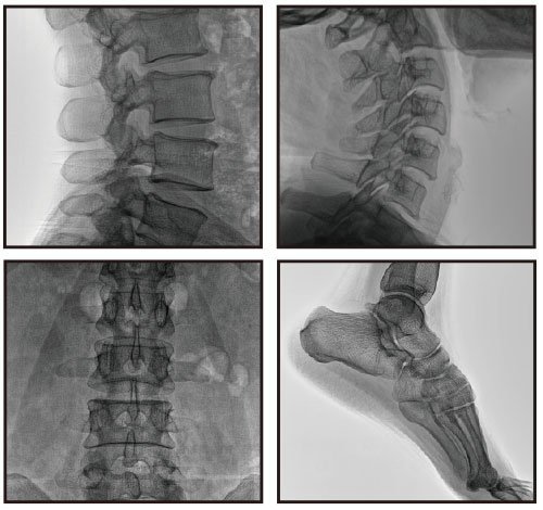
    1. Trauma surgery. For example, in the internal fixation of limb and pelvic fractures, the image is clear and accurate positioning and screw placement can be performed. The simple operation can greatly shorten the fluoroscopy time and reduce the probability of intraoperative and postoperative complications.

    2. Spine and joint surgery. Mainly used in vertebroplasty, elbow, hip, knee joint replacement and anatomical reduction positioning. It can assist in the accurate placement of the prosthesis and reduce the radiation dose.

    3. Obstetrics and gynecology, pain department. For example, recanalization of fallopian tube blockage in gynecology, pain treatment with ozone machine in pain department, etc.; compact frame, easy to exercise, simple to operate, and convenient for doctors to use.


    The above mainly introduces the clinical application and advantages of the mobile C-arm. In fact, as the scope of clinical applications expands, its advantages will become more prominent, and with the support of policies, the market competitiveness of the mobile C-arm will continue to rise. At present, many brands and manufacturers are constantly entering the market, so when choosing, it is necessary to compare more and choose a suitable product. If you are interested in the mobile C-arm products of Perlove Medical,pls contact us soon


What is a 3D C-arm machine?

 What is a 3D C-arm machine? The 3D C-arm machine is a new type of C-arm, which realizes the leap from two-dimensional to three-dimensional intraoperative images. Through the tomographic images of different directions, it can assist the doctor to obtain more comprehensive image information, observe the accuracy of screw implantation, and improve the accuracy of the operation, and the doctor can also check the operation results in time during the operation to confirm the completion of the operation, which greatly improves the accuracy of the operation. Improve the success rate of surgery, reduce the risk of surgery and the probability of postoperative complications.


3D C-arm machine


Puai Medical is a professional manufacturer of C-arm machines. It is the first to master 3D imaging technology in China. Following the first domestic 3D shadow-enhanced C-arm, it has also launched an isocenter flat 3D C-arm machine - Skybow PLX7500. Next, I will introduce the clinical application and advantages of this product of our company.


Clinical application of Skybow PLX7500 3D C-arm machine:


1. Spine Surgery. It can effectively reconstruct the complex three-dimensional model of the spine, provide accurate two-dimensional and three-dimensional images, improve the accuracy of screw implantation, shorten the operation time, reduce the probability of complications, and reduce the intake of radiation.


2. Trauma Surgery. For example, internal fixation of pelvic fractures can reconstruct the acetabular articular surface in three dimensions, accurately locate and implant screws, effectively shorten the time of screw implantation and fluoroscopy exposure, and reduce the probability of postoperative complications.


3. Joint Surgery. It is mainly used in the positioning of joint replacement and anatomical reduction of shoulder joint, elbow joint, hip joint and knee joint. It can accurately implant the prosthesis, reduce the radiation time, and reduce the radiation dose.


4. Osteotomy Orthopaedic Surgery. For example, in the treatment of congenital kyphoscoliosis, it can effectively guide the implantation of screws and remove the half cone to achieve a satisfactory osteotomy correction effect.


Advantages and features of Skybow PLX7500 3D C-arm machine:

1. Intraoperative real-time 3D imaging. Intraoperative three-dimensional imaging and cross-sectional images can provide multi-angle surgical diagnosis information, assisting doctors in intraoperative assessment and judgment, such as fracture reduction and the size and position of implanted screws, and assisting in better completion of surgery.


2. 3D imaging field of view is large. It provides a larger intraoperative 3D imaging field and collects more image information. It can shoot the entire cervical spine, the entire lumbar spine, seven thoracic vertebrae, bilateral sacroiliac joints, femoral head and unilateral pelvis at one time.


3. Isometric design. During the C-arm scanning process, the subject is always kept in the center of the beam, which avoids horizontal and vertical movement during the acquisition of sequential images, and reduces motion artifacts caused by relative movement.


4. Five-dimensional electric operation is convenient. The vertical lifting movement of the plate is convenient for fine-tuning the distance between the plate and the subject during the operation, which is closer to the lesion body, and the imaging range is larger and the image is clearer.


Perlove SKYBOW PLX7500 3D C-arm machine adopts 30cm x 30cm dynamic flat panel detector to output high-resolution, large-size 2D and 3D images. 16-bit grayscale, high dynamic range clearly shows the position and contour of the implant. The imaging is clear, the operation is simple, and the positioning is convenient. It is mainly suitable for orthopedics, spine surgery, orthopedics, trauma orthopedics and operating rooms, etc.